Veterinary medicine has changed more in the past decade than in the half-century that preceded it. The diagnostic tools available to a well-equipped animal hospital today were, until recently, reserved for university teaching facilities and specialty referral centres. Pets across the Greater Toronto Area now benefit from imaging, lab testing, and minimally invasive procedures that catch problems early — sometimes before any visible symptoms appear. At Dixie Animal Hospital Mississauga, the day-to-day reality of modern veterinary care leans heavily on this technology. This guide explains what that actually means for pet owners, what each tool does, and how to evaluate whether a clinic is keeping up.
Why Modern Vet Technology Matters for Pet Outcomes
The single biggest shift in veterinary medicine over the last fifteen years is the move from “wait and see” to “see and act.” Traditional clinics relied heavily on physical exams, basic bloodwork, and educated guesses. Modern clinics combine those foundations with imaging and lab tools that produce answers in minutes rather than days.
This matters more than it sounds. According to the American Veterinary Medical Association, early detection of conditions like kidney disease, heart abnormalities, and certain cancers significantly improves treatment outcomes — and often reduces the total cost of long-term care. A 2024 AAHA diagnostic imaging report found that clinics offering in-house digital imaging and ultrasound detect treatable conditions at earlier stages compared to facilities that send out for the same tests.
Speed is the other variable. When a pet arrives in distress, the difference between getting an X-ray result in two minutes versus two hours can shape the entire treatment path. Animals do not articulate symptoms; their bodies do. The clinics best positioned to listen are the ones with the equipment to translate.
The good news for Mississauga pet owners is that the Dixie veterinary clinic and a handful of others in the region have invested seriously in this tier of equipment, narrowing the gap between general practice and specialty care.
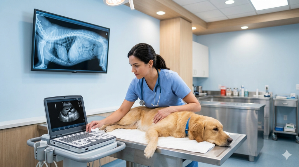
Digital X-rays for Pets: Imaging Without Invasion
Radiography is the workhorse of veterinary diagnostics. Almost every clinic offers it, but the quality varies enormously depending on whether the equipment is analog film, computed radiography, or fully digital.
How Digital Radiography Works
Digital X-rays use direct sensors to capture the image and send it to a computer screen within seconds. There is no film, no chemical processing, and no waiting. The veterinarian can zoom, adjust contrast, and rotate the image to inspect specific regions — capabilities that simply do not exist with traditional film.
Radiation exposure is also lower with modern digital systems. Most clinics using this technology operate at a fraction of the dose required for older equipment, which matters for pets that need repeat imaging and for the staff handling them daily.
What Digital X-rays Reveal
Radiographs are the first-line tool for evaluating:
- Bones and joints — fractures, arthritis, hip dysplasia, and developmental issues
- Chest cavity — heart size, lung patterns, fluid accumulation, and masses
- Abdomen — organ size, foreign object ingestion, bladder stones, and tumours
- Dental health — root abscesses and bone loss invisible during oral inspection
The clinics offering digital radiography on-site can rule in or rule out major problems within the same visit. That speed is the whole point. A pet that swallowed a sock can be diagnosed and prepped for treatment in the same appointment, rather than referred elsewhere and made to wait. For a deeper look at how diagnostic capability factors into clinic quality, this guide on evaluating a trustworthy Dixie vet clinic is worth reading.
Ultrasound Veterinary Diagnostics Explained
If X-rays tell you about bones and density, ultrasound tells you about soft tissue and movement. The two are complementary, not interchangeable. A clinic offering only one is working with half the picture.
Soft Tissue Visualization
Ultrasound uses high-frequency sound waves to produce real-time images of internal structures. Unlike X-rays, there is no radiation, and unlike CT scans, there is no sedation required for most studies. A trained veterinarian can examine the heart, liver, kidneys, spleen, intestines, and reproductive organs while the pet rests quietly on the exam table. The World Small Animal Veterinary Association emphasizes ultrasound as a foundational tool in modern small animal practice, noting how dramatically it has changed primary-care diagnostics.
The technology is particularly useful for cats, who tolerate ultrasound far better than they tolerate restraint for X-rays. It is also the diagnostic of choice for pregnancy confirmation, evaluating abdominal pain, and tracking known conditions over time.
Common Ultrasound Applications
Modern veterinary ultrasound is used routinely for:
- Cardiac assessment — measuring chamber sizes, valve function, and contractility
- Abdominal exploration — checking for fluid, masses, organ changes, or obstructions
- Reproductive health — confirming pregnancy and monitoring fetal development
- Targeted aspirations — guiding needles to specific organs or lesions for sampling
- Soft tissue injuries — assessing tendon, muscle, and ligament damage
A pet ultrasound near me has become a routine expectation rather than a specialist-only service. The Canadian Veterinary Medical Association has noted growth in ultrasound adoption among general practice clinics over the past several years, with most full-service hospitals now offering at least basic abdominal and cardiac scans.
Veterinary Endoscopy: Minimally Invasive Internal Exams
Endoscopy is where modern vet technology becomes genuinely transformative. Instead of opening an animal up surgically to investigate a problem, a veterinarian can use a flexible camera-tipped scope to look directly inside the body — often through natural openings.
How Veterinary Endoscopy Works
A vet endoscope is a long, narrow flexible tube with a high-resolution camera, light source, and working channel for instruments. The veterinarian guides the scope through the mouth, rectum, or a small surgical port and views the internal anatomy on a high-definition monitor in real time.
The procedure typically requires brief general anesthesia, but recovery is dramatically faster than traditional exploratory surgery. Most pets are home the same day, walking comfortably and eating normally within hours.
When Endoscopy Is the Right Choice
Endoscopy is the preferred tool for:
- Removing swallowed foreign objects from the esophagus or stomach without surgery
- Investigating chronic vomiting, diarrhea, or weight loss that bloodwork cannot explain
- Taking precise tissue biopsies from the gastrointestinal tract
- Examining airway issues, chronic coughing, or upper-respiratory abnormalities
A well-trained team can resolve cases in a single endoscopy visit that, twenty years ago, would have meant invasive surgery and a week of recovery. This is the kind of advance that the advanced veterinary diagnostics and treatment capabilities at modern animal hospitals make possible.
In-House Laboratory Testing and Rapid Bloodwork
The fourth pillar of modern diagnostics is what happens after the imaging. Blood, urine, and tissue samples used to be packaged, shipped overnight to a reference lab, and reviewed two to three days later. Clinics with in-house laboratories now run comprehensive panels in fifteen to thirty minutes.
In-house diagnostics typically cover:
- Complete blood counts and chemistry panels for organ function
- Urinalysis for kidney health and infection screening
- Electrolyte and blood gas analysis for critical patients
- Rapid pathogen tests for parvovirus, feline leukemia, and tick-borne diseases
- Cytology for evaluating skin lumps and lymph nodes under the microscope
Reference labs still play an important role for specialized testing — hormone panels, advanced pathology, and culture-based sensitivity testing. But the ability to answer urgent questions immediately, without losing a day or two to shipping, changes how veterinarians make decisions during the appointment itself.
Advanced Pet Diagnostics in Emergency Care
Nowhere does modern equipment matter more than in emergency and urgent care. A pet with sudden collapse, severe vomiting, or suspected toxin exposure does not have time for an external imaging referral.
In a well-equipped emergency setting, a critical patient can be triaged, imaged with digital X-ray and ultrasound, sampled for in-house bloodwork, and started on stabilizing treatment within an hour of arrival. The veterinarian is making decisions based on actual data, not estimates. That difference is often the difference between a successful outcome and a heartbreaking one.
The same equipment that supports emergency cases also supports the more nuanced work of senior pet care services, where age-related conditions develop quietly and need to be caught through routine screening rather than during a crisis.

Case Study: How Imaging Caught What a Routine Exam Missed
A Mississauga family brought their seven-year-old Labrador mix in for what they assumed was a minor issue. The dog had been eating less for about a week, and seemed slightly less energetic on walks. There was no fever, no vomiting, no obvious pain. A traditional exam might have ended with a recommendation to “watch and see.”
Instead, the veterinarian recommended an abdominal ultrasound based on subtle changes felt during palpation. The scan revealed a small splenic mass — the kind that ruptures suddenly and causes life-threatening internal bleeding without warning. Within forty-eight hours, the dog was in surgery for a successful splenectomy. The mass turned out to be benign.
The owners later said they would have waited weeks before pushing for further investigation if the clinic had not had the imaging capability on-site. That early detection, made possible by in-house ultrasound, is exactly the scenario advanced diagnostic technology is designed for. Online reviews of the animal hospital Mississauga consistently echo this theme — the team finds things others miss, and explains the findings clearly enough that owners can make informed decisions.
Comparison: Traditional vs. Modern Vet Technology
| Capability | Traditional Clinic | Modern Equipped Clinic |
|---|---|---|
| X-ray imaging | Film, slow processing | Digital, results in seconds |
| Soft tissue imaging | Referral to specialist | On-site ultrasound |
| Internal investigation | Exploratory surgery | Minimally invasive endoscopy |
| Bloodwork turnaround | 2–3 days via reference lab | 15–30 minutes in-house |
| Image sharing | Physical films couriered | Digital files emailed instantly |
| Emergency triage | Limited on-site data | Full diagnostic workup |
| Records storage | Paper charts | Cloud-based, searchable |
Frequently Asked Questions
-
Is digital X-ray safer than traditional film X-ray for pets?
Yes. Digital radiography systems typically use significantly lower radiation doses than older film-based equipment to produce a clear image. The combination of lower exposure and the ability to retake misaligned images instantly — without re-dosing the pet — makes digital imaging both safer and more accurate. Most modern Mississauga clinics have transitioned fully to digital. If a clinic still relies on film, that is a meaningful signal about their overall equipment investment.
-
When does a pet need an ultrasound instead of an X-ray?
The two tools answer different questions. X-rays excel at evaluating bones, the chest cavity, and dense structures. Ultrasound is the better choice for soft tissue — organs, fluid, masses, and blood flow patterns. Many serious conditions require both. A veterinarian will recommend one, the other, or both based on the suspected problem. Clinics offering both on-site can complete a full workup in one visit, which matters during time-sensitive situations.
-
Is endoscopy painful for pets?
Endoscopy itself is not painful — pets are under general anesthesia for the procedure and feel nothing during it. Mild throat irritation or temporary fatigue is possible afterward, depending on the type of scope used and how long the procedure took. Most pets return to normal eating and activity within twenty-four hours. Compared to traditional exploratory surgery, recovery is dramatically faster and less uncomfortable, which is why endoscopy is now preferred whenever clinically appropriate.
-
How much does advanced veterinary diagnostic equipment add to my pet’s visit?
The cost of individual diagnostics depends on the procedure, but the broader picture matters more. Catching a treatable condition early with imaging routinely saves owners from far more expensive emergency surgery or long-term management of advanced disease. Discuss expected costs transparently with the clinic before procedures begin. Trustworthy teams will explain why a specific test is recommended and what the results will or will not tell them about your pet’s situation.
-
How do I find a vet clinic with modern diagnostic technology?
Look for clinics that publish their equipment list on their website, mention digital radiography, ultrasound, and in-house laboratory capability, and welcome questions about their tools. Reviews often mention specific scenarios where technology made a difference. A practical option is to call ahead and ask whether they perform same-visit imaging and bloodwork. To explore one full-service option, the neighbourhood pet clinic guide outlines what to look for in detail. You can also reach out directly through their veterinary care near me contact page to ask specific questions before booking.
The right diagnostic technology in the hands of an attentive veterinary team is what turns guesswork into precise care — and that, ultimately, is the standard every pet deserves.